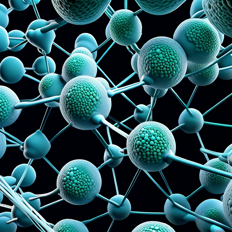
【what is shiba inu coin worth right now】Tether Protein Configuration: Exploring the Architecture of Cellular Anchor Points
This article delves into the fascinating world of tether protein structures,what is shiba inu coin worth right now their roles within cellular mechanisms, and the significance they hold for understanding complex biological processes. Tether proteins serve as crucial components in the architectural and functional layout of cells, playing an instrumental role in intracellular transport, membrane fusion events, and the maintenance of organelle identity. By dissecting their structure, one can uncover the intricate ways in which these proteins contribute to cellular dynamics and overall organismal health.


The Fundamental Structure of Tether Proteins
At the most fundamental level, tether proteins are characterized by their ability to anchor various cellular components to specific locations within the cell. This anchoring mechanism is vital for maintaining the spatial organization of cellular organelles and for facilitating the targeted delivery of vesicles during processes such as exocytosis and endocytosis. Structurally, tether proteins are diverse, but they often contain coiled-coil domains or lipid-binding motifs that enable them to interact with membrane surfaces and other proteins. This structural variability allows tether proteins to perform a wide range of functions, adapting to the specific requirements of the cellular compartments they serve.
Roles and Functions in Intracellular Transport
One of the primary roles of tether proteins is in the mechanism of intracellular transport. They act as molecular bridges that facilitate the movement of vesicles between different compartments within the cell. Through their specific structural features, tether proteins can recognize and bind to vesicles, guiding them to their target destinations. This precise guidance is crucial for the efficiency of vesicular transport systems and is instrumental in processes such as neurotransmitter release, hormone secretion, and lysosomal degradation. By understanding the structural basis of tether proteins’ involvement in intracellular transport, scientists can gain insight into how cellular materials are trafficked with such specificity and speed.
Contribution to Membrane Fusion Events
Beyond their role in transport, tether proteins are also key players in the mechanism of membrane fusion. Fusion events are essential for various cellular processes, including vesicle docking and the release of their contents into specific cellular locations. Tether proteins contribute to these events by initially recognizing and attaching to the target membrane, bringing the vesicle membrane into close proximity. This action facilitates the merging of lipid bilayers, a critical step towards the successful delivery of vesicle contents. The structure of tether proteins provides the necessary specificity and affinity for interacting with distinct membrane lipids or proteins, ensuring that fusion occurs at the correct spatial and temporal location.
In conclusion, tether proteins play an indispensable role in the orchestration of cellular activities through their versatile structural features. Their ability to anchor, guide, and merge cellular components is fundamental to intracellular transport, organelle organization, and the execution of precise cellular mechanisms. The study of tether protein structures not only sheds light on their essential functions within the cell but also offers broader insights into the complexities of biological systems and potential therapeutic targets for addressing cellular dysfunction.